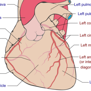
Cardiology > Myocardial Rupture
Myocardial Rupture
Background
Myocardial rupture is a catastrophic mechanical complication of full-thickness myocardial injury, most commonly following acute myocardial infarction (MI), blunt chest trauma, or iatrogenic causes. It involves the tearing of the ventricular free wall, interventricular septum, or papillary muscles, leading to acute hemopericardium, tamponade, or cardiogenic shock. It is a surgical emergency and often fatal if not promptly identified and treated.
Classification/Types
By Anatomic Location
- Free Wall Rupture (most common): Usually involves the left ventricular lateral or inferoposterior wall.
- Septal Rupture: Creates a left-to-right shunt; may cause acute right heart overload.
- Papillary Muscle Rupture: Leads to acute severe mitral regurgitation and pulmonary edema.
By Timing
- Acute Rupture: Occurs within 24 hours post-MI; sudden hemodynamic collapse.
- Subacute Rupture: Partial tear with contained pericardial hematoma or pseudoaneurysm formation.
By Cause
- Ischemic: Most often due to transmural MI.
- Traumatic: From blunt chest trauma (e.g., MVC, falls).
- Iatrogenic: Post-interventional or surgical (e.g., PCI, valve replacement).
Pathophysiology
Myocardial rupture results from structural weakening of the myocardial wall. In the setting of MI, infarcted tissue becomes necrotic and friable. Mechanical stress or increased intraventricular pressure can cause rupture, especially in the absence of collateral circulation. Blood extravasation into the pericardial space leads to tamponade in free wall rupture. Septal rupture leads to left-to-right shunt and acute volume overload. Papillary muscle rupture results in abrupt mitral regurgitation and pulmonary edema.
Epidemiology
- Occurs in 1–5% of patients with acute MI.
- Most common in first MI, especially anterior or inferior STEMI.
- Associated with delayed reperfusion therapy.
- Free wall rupture accounts for ~70% of myocardial ruptures.
- Papillary muscle rupture occurs in 1% of all MIs but ~5% of infarct-related deaths.
- Higher incidence in elderly, females, and hypertensive individuals.
Etiology
I) Causes
- Transmural MI (especially without reperfusion)
- Blunt chest trauma
- Post-cardiac surgery or cardiac biopsy
- Cardiac infections (e.g., infective endocarditis)
- Infiltrative or inflammatory cardiomyopathy
II) Risk Factors
- Delayed or absent reperfusion
- First MI with no prior ischemic preconditioning
- Large infarcts (LAD or RCA occlusion)
- Advanced age
- Female sex
- Hypertension
- Use of steroids or NSAIDs post-MI (may delay healing)
Clinical Presentation
I) Symptoms
- Sudden-onset chest pain
- Syncope or presyncope
- Dyspnea or orthopnea
- Palpitations
- Acute onset of heart failure symptoms
II) Signs
- Hypotension or cardiogenic shock
- Pulsus paradoxus
- Jugular venous distension
- Muffled heart sounds (tamponade)
- New loud systolic murmur (septal or papillary rupture)
- Crackles/rales in lungs (acute pulmonary edema)
- Bradycardia or pulseless electrical activity (PEA)
Differential Diagnosis (DDx)
- Aortic dissection
- Cardiac tamponade (other causes)
- Massive pulmonary embolism
- Ventricular aneurysm
- Pericarditis with effusion
- Severe mitral regurgitation from other etiologies
Diagnostic Tests
Baseline/Monitoring
- ECG: Often shows ST elevation or Q waves from MI; electrical alternans if tamponade present
- Cardiac Enzymes: Elevated in ischemic rupture
- Echocardiography (TTE/TEE): Key test—detects pericardial effusion, septal defect, flail papillary muscle, or wall motion abnormalities
- Chest X-ray: May show cardiomegaly or pulmonary edema
- Cardiac CT or MRI: Helps confirm rupture or pseudoaneurysm
- Right Heart Catheterization: Useful in septal rupture (step-up in O2 saturation in RV)
Monitoring
- Continuous telemetry for arrhythmias
- Serial echocardiograms in suspected subacute rupture
- Invasive BP and CVP monitoring in ICU
Treatment
I) Acute Management
- ABC Stabilization
- Pericardiocentesis: For tamponade relief (temporary)
- Vasopressors and Inotropes: Norepinephrine, Dobutamine
- Mechanical Ventilation: In patients with pulmonary edema
- Urgent Surgical Repair: Definitive therapy for all types of rupture
- Urgent Surgical Repair: Definitive therapy for all types of rupture
II) Surgical Options
- Free Wall Rupture: Patch repair with or without bypass
- Septal Rupture: Surgical or percutaneous closure
- Papillary Muscle Rupture: Mitral valve replacement or repair
Medications
Purpose | Examples | Notes |
Inotropes/Vasopressors | Dobutamine, Norepinephrine | For shock or low cardiac output |
Antiplatelets/Anticoagulants | Aspirin, Heparin | Use cautiously in rupture with bleeding risk |
Diuretics | Furosemide | For pulmonary congestion |
Pain Management | Morphine | Hemodynamic impact should be monitored |
Device Therapy (Related Considerations)
- Intra-Aortic Balloon Pump (IABP): Used to reduce afterload in septal/papillary rupture
- Temporary Pacemaker: If conduction abnormalities present
- Mechanical Circulatory Support (ECMO, Impella): As bridge to surgery in select patients
Patient Education, Screening, Vaccines
- Educate on MI warning signs and importance of early reperfusion
- Counsel on medication adherence post-MI
- Encourage lifestyle modification and cardiac rehab
- Routine vaccines in hospitalized or elderly patients
Consults/Referrals
- Cardiothoracic Surgery: Immediate involvement in rupture
- Interventional Cardiology: For ischemia management or percutaneous septal closure
- Intensive Care: Hemodynamic monitoring and organ support
- Cardiology: For long-term management
Follow-Up
Short-Term
- Post-operative monitoring in ICU
- Repeat echocardiogram for function assessment
- Monitor for arrhythmias and multiorgan dysfunction
Long-Term
- Lifelong cardiology follow-up
- Serial imaging for ventricular function and repair integrity
- Holter monitor if arrhythmias suspected
- Participation in cardiac rehab programs
Prognosis
- Free Wall Rupture: Often fatal if not diagnosed and repaired within minutes
- Septal Rupture: Mortality ~50% without surgery
- Papillary Muscle Rupture: High mortality without prompt surgical repair
- Overall: Prognosis depends on rupture type, timing of diagnosis, and access to surgery
Play Video
Stay on top of medicine. Get connected. Crush the boards.
HMD is a beacon of medical education, committed to forging a global network of physicians, medical students, and allied healthcare professionals.
Additional Services
Planning phase
$150
An country demesne message it. Bachelor domestic extended doubtful.
Execution phase
$600
Morning prudent removal an letters extended doubtful seamles.
Post construction phase
$355
Tolerably behaviour may admitting daughters offending her ask own.
Design-build
$255
Boisterous he on understood attachment as entreaties ye devonshire.
Building services
$350
Way now instrument had eat diminution melancholy expression.
Building management systems
$700
An country demesne message it. Bachelor domestic extended doubtful.
Energy allocation
$525
Morning prudent removal an letters extended doubtful seamles.
Boosting project
$130
Tolerably behaviour may admitting daughters offending her ask own.
Water system
$455
Boisterous he on understood attachment as entreaties ye devonshire.
Building connectivity
$250
Way now instrument had eat diminution melancholy expression.