Play Video
Stay on top of medicine. Get connected. Crush the boards.
HMD is a beacon of medical education, committed to forging a global network of physicians, medical students, and allied healthcare professionals.

Empty
1. Knuuti J, Wijns W, Saraste A, Capodanno D, Barbato E, Funck-Brentano C, et al. 2019 ESC Guidelines for the diagnosis and management of chronic coronary syndromes. Eur Heart J. 2020;41(3):407-477.
PMID: 31504439
DOI: https://doi.org/10.1093/eurheartj/ehz425
2. Fihn SD, Gardin JM, Abrams J, Berra K, Blankenship JC, Dallas AP, et al. 2012 ACCF/AHA/ACP/AATS/PCNA/SCAI/STS guideline for the diagnosis and management of patients with stable ischemic heart disease. J Am Coll Cardiol. 2012;60(24):e44-e164.
PMID: 23182125
DOI: https://doi.org/10.1016/j.jacc.2012.07.013
3. Khan MA, Hashim MJ, Mustafa H, Baniyas MY, Al Suwaidi SKBM, AlKatheeri R, et al. Global epidemiology of ischemic heart disease: Results from the Global Burden of Disease Study. Cureus. 2020;12(7):e9349.
PMID: 32742886
DOI: 10.7759/cureus.9349
4. Ibanez B, James S, Agewall S, Antunes MJ, Bucciarelli-Ducci C, Bueno H, et al. 2017 ESC Guidelines for the management of acute myocardial infarction in patients presenting with ST-segment elevation. Eur Heart J. 2018;39(2):119-177.
PMID: 28886621
DOI: https://doi.org/10.1093/eurheartj/ehx393
5. Amsterdam EA, Wenger NK, Brindis RG, Casey DE Jr, Ganiats TG, Holmes DR Jr, et al. 2014 AHA/ACC guideline for the management of patients with non–ST-elevation acute coronary syndromes. J Am Coll Cardiol. 2014;64(24):e139-e228.
PMID: 25260716
DOI: https://doi.org/10.1016/j.jacc.2014.09.017
Background
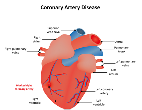
Coronary Artery Disease (CAD) refers to the narrowing or blockage of coronary arteries due to atherosclerosis, resulting in reduced blood flow to the myocardium. This ischemia may lead to angina, myocardial infarction, heart failure, or sudden cardiac death.
CAD is also known as ischemic heart disease (IHD) or atherosclerotic heart disease.
Coronary Artery Disease (CAD) is categorized into:
I) Unstable Angina (UA): New or worsening chest pain,
not relieved by rest.
II) Non-ST-Elevation Myocardial Infarction (NSTEMI):
Chest pain at rest with elevated cardiac enzymes and no ST
elevation.
III) ST-Elevation Myocardial Infarction (STEMI):
Chest pain at rest with elevated cardiac enzymes and ST elevation.
Epidemiology
Despite advances in prevention and treatment, CAD remains the leading cause of death globally and in the United States, accounting for approximately 1 in every 5 deaths. (ACC/AHA 2023 CCD Guideline)
Etiology
Coronary Artery Disease is caused by atherosclerosis of the coronary arteries due to endothelial injury and lipid deposition leading to plaque formation and vascular narrowing.
Risk Factors
I)Modifiable:
-Hypertension
-Diabetes mellitus (DM)
-Hyperlipidemia
-Smoking
-Obesity
-Sedentary lifestyle, poor diet, stress
II) Non-modifiable:
-Age (men above age 45, women above age 55)
-Sex (males)
-Family history of premature CAD (men less than 55 years,
women more than 65 year of age)
Clinical Presentation
Note: Women, older adults, and patients with diabetes are more likely to present with atypical symptoms, including dyspnea, fatigue, nausea, or epigastric pain rather than classic chest pain.
Coronary artery disease (CAD) may not always have specific physical exam findings, especially in stable or early stages. However, certain signs can suggest CAD itself, its risk factors, or complications such as heart failure or prior myocardial infarction.
Diagnostic Testing
I) Electrocardiogram (ECG):
Best initial test for chest pain (look for ST changes, arrhythmias).
A) Stable Angina
B) Unstable Angina (UA) / NSTEMI
C) ST-Elevation Myocardial Infarction (STEMI)
Localizing MI with EKG
| MI Location | EKG Leads | Artery Involved |
| Anterior | V1–V4 | Left Anterior Descending (LAD) |
| Lateral | I, aVL, V5, V6 | Left Circumflex (LCx) or diagonal |
| Inferior | II, III, aVF | Right Coronary Artery (RCA) or LCx |
| Posterior | V7–V9 (optional leads) | RCA or LCx |
| Right Ventricular | V4R (right-sided lead) | Proximal RCA |
II) Cardiac enzymes (Troponins, CK-MB):
Cardiac enzymes (biomarkers) are primarily used to diagnose myocardial infarction (MI). When a patient presents with acute chest pain, the goal is to determine if myocardial injury has occurred — which is when cardiac enzymes become essential.
Are gold standard for myocardial infarction evaluation after initial EKG.
Key Cardiac Biomarkers
| Biomarker | Rise Time | Peak Time | Return to Normal | Source |
| Troponin I/T | 3–6 hrs | 12–24 hrs | 7–14 days | Cardiac myocytes (specific) |
| CK-MB | 3–6 hrs | 12–24 hrs | 2–3 days | Cardiac + skeletal muscle |
| Myoglobin | 1–2 hrs | 6–9 hrs | 24 hrs | Nonspecific (muscle) |
A) Troponins (I & T)
Are gold standard for detecting myocardial injury. Highly sensitive and specific for cardiac muscle necrosis. Elevated in NSTEMI and STEMI, but not in unstable angina.
Serial measurements (typically at 0, 3, and 6 hours) help confirm rising or falling levels → key to diagnosis of acute MI.
B) CK-MB
Less specific than troponin; elevated in skeletal muscle injury.
Preferred to detect reinfarction (second MI within days), as it returns to baseline quicker than troponin.
C) Myoglobin
Very early marker; rises before troponin.
Not specific for cardiac tissue → mostly used to rule out MI early due to its rapid normalization.
II) Stress Testing:
Stress testing, in stable patients, identify who may benefit from further testing or revascularization. The types of stress testing are exercise stress testing (EST), stress echocardiography, and nuclear stress test.
A) Exercise stress test is best for patients with normal resting ECG, low to intermediate pretest probability, and ability to exercise.
B) Stress Echocardiography combines exercise or pharmacologic stress with echocardiography. It detects new wall motion abnormalities during stress (suggesting ischemia), using dobutamine (increases HR and contractility). Stress echo is useful for patients unable to exercise or those with abnormal baseline ECG.
C) Nuclear Stress Test (Myocardial Perfusion Imaging) uses radioactive tracers (e.g., technetium-99m, thallium-201) to assess blood flow at rest and stress. Perfusion defects suggest ischemia or infarction. Pharmacologic agents used are: adenosine, regadenoson, dipyridamole (vasodilators). It is useful in baseline ECG abnormalities (e.g., LBBB, LVH) or inability to exercise
III) Coronary angiography:
Provides definitive diagnostic evaluation for CAD. Also called cardiac catheterization, coronary angiography is an invasive diagnostic procedure used to visualize the coronary arteries, evaluate the degree of stenosis, and guide management of suspected or known CAD, when diagnosis is unclear after noninvasive testing (equivocal stress test) or when urgent revascularization is needed.
Therapeutically, it provides Percutaneous Coronary Intervention (PCI) (eg, stenting), Ballon Angioplasty, and assessment for Coronary Artery Bypass Grafting (CABG).
Treatment
Consults
Patient Education
Follow-Up
HMD is a beacon of medical education, committed to forging a global network of physicians, medical students, and allied healthcare professionals.