Medicine, via pristina

Medicine, via pristina

Ventricular Fibrillation

1. Knuuti J, Wijns W, Saraste A, Capodanno D, Barbato E, Funck-Brentano C, et al. 2019 ESC Guidelines for the diagnosis and management of chronic coronary syndromes. Eur Heart J. 2020;41(3):407-477.
PMID: 31504439
DOI: https://doi.org/10.1093/eurheartj/ehz425


2. Fihn SD, Gardin JM, Abrams J, Berra K, Blankenship JC, Dallas AP, et al. 2012 ACCF/AHA/ACP/AATS/PCNA/SCAI/STS guideline for the diagnosis and management of patients with stable ischemic heart disease. J Am Coll Cardiol. 2012;60(24):e44-e164.
PMID: 23182125
DOI: https://doi.org/10.1016/j.jacc.2012.07.013


3. Khan MA, Hashim MJ, Mustafa H, Baniyas MY, Al Suwaidi SKBM, AlKatheeri R, et al. Global epidemiology of ischemic heart disease: Results from the Global Burden of Disease Study. Cureus. 2020;12(7):e9349.
PMID: 32742886
DOI: 10.7759/cureus.9349


4. Ibanez B, James S, Agewall S, Antunes MJ, Bucciarelli-Ducci C, Bueno H, et al. 2017 ESC Guidelines for the management of acute myocardial infarction in patients presenting with ST-segment elevation. Eur Heart J. 2018;39(2):119-177.
PMID: 28886621
DOI: https://doi.org/10.1093/eurheartj/ehx393


5. Amsterdam EA, Wenger NK, Brindis RG, Casey DE Jr, Ganiats TG, Holmes DR Jr, et al. 2014 AHA/ACC guideline for the management of patients with non–ST-elevation acute coronary syndromes. J Am Coll Cardiol. 2014;64(24):e139-e228.
PMID: 25260716
DOI: https://doi.org/10.1016/j.jacc.2014.09.017

Background 

I) Definition 

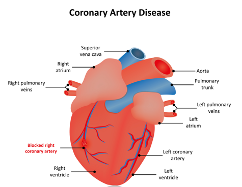
Ventricular fibrillation (VF) is a life-threatening arrhythmia characterized by rapid, chaotic, and disorganized electrical activity in the ventricles, resulting in ineffective myocardial contraction and immediate cessation of cardiac output. VF is a form of pulseless cardiac arrest and the most common arrhythmia associated with sudden cardiac death (SCD). Without rapid defibrillation, it is uniformly fatal. 

I) Classification/Types 

By Duration: 

  • Primary VF: Occurs within the first hour of acute myocardial infarction (AMI), not preceded by hemodynamic deterioration. 
  • Secondary VF: Occurs in response to profound ischemia or metabolic derangement, often with preceding hemodynamic compromise. 

By Association: 

  • Ischemic VF: Commonly related to acute coronary syndromes 
  • Non-ischemic VF: Due to structural heart disease, electrolyte imbalance, or inherited arrhythmias 
  • Idiopathic VF: No identifiable cause, often suspected in survivors of unexplained cardiac arrest 


Pathophysiology 

VF arises from multiple, disorganized reentrant wavelets of electrical activity that circulate within the ventricular myocardium. This disorganized excitation prevents coordinated contraction, leading to no effective cardiac output. Common initiating factors include ischemia-induced dispersion of repolarization, reentrant circuits in scarred myocardium, or early afterdepolarizations (especially in long QT syndromes). Without rapid intervention, VF progresses to asystole


Epidemiology 

  • VF is the leading cause of sudden cardiac death (SCD) globally 
  • Occurs most commonly in adult males >45 years, especially those with coronary artery disease 
  • Major cause of out-of-hospital cardiac arrest 
  • Rare in children unless due to congenital cardiac or electrical disorders 


Etiology
 

I) Causes 

  • Acute myocardial infarction (most common) 
  • Cardiomyopathies (dilated, hypertrophic) 
  • Electrolyte abnormalities (hypokalemia, hypomagnesemia) 
  • Drug toxicity (e.g., digoxin, QT-prolonging agents) 
  • Inherited channelopathies: Long QT syndrome, Brugada syndrome, catecholaminergic polymorphic VT 
  • Severe heart failure 
  • Commotio cordis (blunt chest trauma during vulnerable repolarization phase) 

II) Risk Factors 

  • History of coronary artery disease or MI 
  • Prior episode of VT or VF 
  • Reduced left ventricular ejection fraction (LVEF) 
  • Family history of sudden cardiac death 
  • Use of QT-prolonging drugs 
  • Electrolyte imbalance or renal dysfunction 


Clinical Presentation
 

I) History (Symptoms) 

  • Sudden collapse (primary presentation) 
  • Witnessed syncope or cardiac arrest 
  • Preceding symptoms may include palpitations, chest pain, lightheadedness, or dyspnea (rarely lasts more than seconds before arrest) 
  • No pulse or respiration at time of presentation 

II) Physical Exam (Signs) 

  • Unresponsiveness 
  • Pulselessness 
  • Apnea 
  • Cyanosis and cardiac arrest without preceding signs in most cases 


Differential Diagnosis (DDx)
 

  • Pulseless VT 
  • Asystole 
  • Pulseless electrical activity (PEA) 
  • Torsades de pointes (form of polymorphic VT) 
  • High-grade AV block with bradyasystole 
  • Massive pulmonary embolism or cardiac tamponade (nonarrhythmic arrest) 


Diagnostic Tests
 

Initial Work-Up (in resuscitated patients or survivors) 

  • ECG (post-resuscitation): ST-elevation or Q waves suggest MI 
  • Cardiac enzymes: Assess for myocardial injury 
  • Electrolytes and renal function: Evaluate for correctable triggers 
  • Toxicology screen: Rule out drug-induced causes 
  • Echocardiography: Assess for structural disease 
  • Coronary angiography: If ACS suspected 
  • Cardiac MRI: Evaluate for scar, myocarditis, or cardiomyopathy 
  • Genetic testing: If inherited arrhythmia suspected 
  • EPS: In select cases for arrhythmia source identification 


Treatment
 

I) Initial Approach (ACLS protocol) 

  • Immediate CPR 
  • Early defibrillation (biphasic: 120–200 J; monophasic: 360 J) 
  • Epinephrine 1 mg IV every 3–5 minutes 
  • Amiodarone 300 mg IV bolus (repeat 150 mg if needed) 
  • Treat reversible causes (5 Hs and 5 Ts) 


II)Post-Resuscitation Management
 

  • Therapeutic hypothermia (targeted temperature management if comatose) 
  • Hemodynamic support (vasopressors/inotropes) 
  • Coronary revascularization if ACS is identified 
  • ICD placement for secondary prevention 
  • Beta-blockers and antiarrhythmics to prevent recurrence 


Medications 

Drug Class 

Examples 

Notes 

Antiarrhythmics 

Amiodarone, Lidocaine 

Used acutely post-VF; amiodarone preferred 

Beta-blockers 

Metoprolol, Carvedilol 

Reduce sympathetic tone and arrhythmic risk 

ACE inhibitors/ARBs 

Lisinopril, Losartan 

Mortality benefit in structural heart disease 

Magnesium sulfate 

IV 

For torsades de pointes or suspected deficiency 


Device Therapy 

  • Implantable Cardioverter-Defibrillator (ICD): Gold standard for survivors of VF or those at high risk (EF ≤35%, prior VT/VF) 
  • Wearable Cardioverter-Defibrillator (WCD): Temporary option while awaiting further evaluation 


Consults/Referrals
 

  • Cardiology: For long-term rhythm management 
  • Electrophysiology: For ICD evaluation and ablation if needed 
  • Emergency Medicine: For acute arrest response 
  • Genetics: If inherited arrhythmia suspected 
  • Critical Care: For post-resuscitation care 


Patient Education, Screening, Vaccines,
 Education 

  • Recognize warning signs of arrhythmias (palpitations, dizziness) 
  • Understand ICD use and what to expect with shocks 
  • Importance of medication adherence and avoiding triggers 
  • CPR training for family members 

Screening/Prevention 

  • ECG or Holter in high-risk individuals 
  • Family screening if genetic arrhythmia suspected 
  • Avoid QT-prolonging drugs in susceptible patients 

Vaccinations 

  • Influenza and pneumococcal vaccines recommended for patients with structural heart disease or ICD 
  • No VF-specific vaccines 


Prognosis 

  • VF without prompt defibrillation: 100% fatal 
  • Survival with good neurologic outcome possible if CPR and defibrillation occur within minutes 
  • ICD therapy reduces risk of sudden cardiac death 
  • Long-term prognosis depends on underlying cause and management 
  • Recurrence risk high without ICD or addressing etiology 


Play Video

Stay on top of medicine. Get connected. Crush the boards.

HMD is a beacon of medical education, committed to forging a global network of physicians, medical students, and allied healthcare professionals.

Additional Services

Planning phase $150
An country demesne message it. Bachelor domestic extended doubtful.
Execution phase $600
Morning prudent removal an letters extended doubtful seamles.
Post construction phase $355
Tolerably behaviour may admitting daughters offending her ask own.
Design-build $255
Boisterous he on understood attachment as entreaties ye devonshire.
Building services $350
Way now instrument had eat diminution melancholy expression.
Building management systems $700
An country demesne message it. Bachelor domestic extended doubtful.
Energy allocation $525
Morning prudent removal an letters extended doubtful seamles.
Boosting project $130
Tolerably behaviour may admitting daughters offending her ask own.
Water system $455
Boisterous he on understood attachment as entreaties ye devonshire.
Building connectivity $250
Way now instrument had eat diminution melancholy expression.
Shopping Basket